Home
/ Pelvic Anatomy Ligaments : Pelvis Boundaries Springerlink : The pelvis is held together by three principal ligaments:
Pelvic Anatomy Ligaments : Pelvis Boundaries Springerlink : The pelvis is held together by three principal ligaments:
Pelvic Anatomy Ligaments : Pelvis Boundaries Springerlink : The pelvis is held together by three principal ligaments:. The pelvis's frame is made up of the bones of the pelvis, which connect the axial skeleton to the femurs, and therefore acts in weight bearing of the upper body. The pelvis is held together by three principal ligaments: Also, the compartmental anatomy of the female pelvis is explained, including the extraperitoneal pelvic spaces. There are two major groups of ligaments that provide nearly all the structure of the pelvis. The femoral ligaments act to stabilize the ball and socket joint of the hip, connecting to the ilium and the ischium.
The pelvic girdle, also known as the hip bone, is composed of three fused bones: The pelvis itself is a paired composite structure made up by three bones (ilium, ischium and pubis) that articulates with the sacral part of the axial spine. Other ligaments attached to bony pelvis include the sacrococcygeal ligaments, pubic symphysis ligaments, and endopelvic fascia ligament. The pelvic girdle (hip girdle) is formed by a single bone, the hip bone or coxal bone (coxal = hip), which serves as the attachment point for each lower limb. The suspensory ligament of the ovary, also infundibulopelvic ligament (commonly abbreviated ip ligament or simply ip ), is a fold of peritoneum that extends out from the ovary to the wall of the pelvis.
Pdf Contemporary Views On Female Pelvic Anatomy from www.researchgate.net Inherent stability of the pelvis is provided by ligaments. Imaios and selected third parties, use cookies or similar technologies, in particular for audience measurement. The pelvic ligaments are strong, thick bands of fibrous tissue that connect the pelvic bones. It extends from the lateral pelvic walls on both sides, and folds over the internal female genitalia, covering their surface anteriorly and posteriorly. This image shows the posterior back view of the female pelvic brim (the bones and ligaments that forms the pelvic region in the female) showing: The broad ligament folds over the fallopian tubes and ovaries and covers them anteriorly and posteriorly. The broad ligament can be further divided into three components. The plexus gives off the anococcygeal nerve, which pierces the sacrotuberous ligament and supplies the skin in the region of the coccyx.
The pelvis is a boney structure at the base of the lumbar spine.
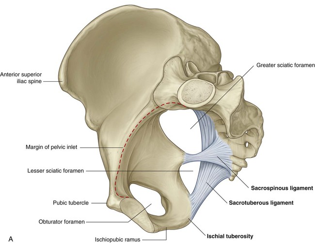
It extends from the lateral pelvic walls on both sides, and folds over the internal female genitalia, covering their surface anteriorly and posteriorly. The cardinal ligaments, also known as the transverse cervical ligaments, the lateral cervical ligaments, or mackenrodt's ligaments, are fibrous bands that attached the cervix to the lateral pelvic walls. This is part of the forced closure method that the pelvis adopts in order to keep itself secure. Iliolumbar, sacrotuberous and sacrospinous ligaments. The broad ligament is a sheet of pelvic peritoneum extending bilaterally from the lateral pelvic sidewalls to the uterus in the midline. Imaios and selected third parties, use cookies or similar technologies, in particular for audience measurement. Bones and ligaments of the female pelvis. Other ligaments attached to bony pelvis include the sacrococcygeal ligaments, pubic symphysis ligaments, and endopelvic fascia ligament. Finally, a checklist is provided for structured reporting of the mri findings in the female pelvis. Clinical anatomy the lumbosacral trunk (l4, l5 ) and the ventral ramus of nerve s1 cross the nerves of the pelvis surface of the joint and may be involved in the disease of the joints, causing pain in the area. There are two major groups of ligaments that provide nearly all the structure of the pelvis. The pelvis is a boney structure at the base of the lumbar spine. The broad ligament can be further divided into three components.
Ligaments connect one bone to another and provide important stability. The broad ligament is related to many structures within the female pelvis. Ligaments and anatomy important in pelvic. The ilium, ischium and the pubic bone. Those that connect the ilium to the sacrum;
Pelvis And Perineum Clinical Gate from clinicalgate.com This image shows the posterior back view of the female pelvic brim (the bones and ligaments that forms the pelvic region in the female) showing: They form what can be described as a basket weave formation, in order to create strength and tensegrity within the structure. It extends from the lateral pelvic walls on both sides, and folds over the internal female genitalia, covering their surface anteriorly and posteriorly. The ligaments that pass between the sacrum and the ischium, which is the lower rear part of the pelvis; The femoral ligaments act to stabilize the ball and socket joint of the hip, connecting to the ilium and the ischium. Ligaments connect one bone to another and provide important stability. We are developing an accurate 3d model of human anatomy. It is usually divided into two separate anatomic regions:
The pelvis is a boney structure at the base of the lumbar spine.
The pelvic girdle, also known as the hip bone, is composed of three fused bones: The ligaments that connect the sacrum to the coccyx. Inherent stability of the pelvis is provided by ligaments. The pelvis consists of two innominate bones and the sacrum to which coccyx is attached. It is usually divided into two separate anatomic regions: The joints of the pelvis are the sacroiliac and sacrococcygeal joints and the pubic symphysis, while the anterior sacroiliac ligament is a flat band which joins the bones above and below the pelvic brim. The broad ligament can be further divided into three components. Clinical anatomy the lumbosacral trunk (l4, l5 ) and the ventral ramus of nerve s1 cross the nerves of the pelvis surface of the joint and may be involved in the disease of the joints, causing pain in the area. The uterosacral ligament supports the uterus posteriorly, and the pubocervical ligament anchors the uterus anteriorly. The ligaments that pass between the sacrum and the ischium, which is the lower rear part of the pelvis; It extends to both sides of the pelvic wall. The suspensory ligament of the ovary (not labeled) is shown incompletely and in section; We are developing an accurate 3d model of human anatomy.
Cookies allow us to analyze and store information such as the characteristics of your device as well as certain personal data (e.g., ip addresses, navigation, usage or geolocation data, unique identifiers). The pelvis is a boney structure at the base of the lumbar spine. The broad ligament is a sheet of pelvic peritoneum extending bilaterally from the lateral pelvic sidewalls to the uterus in the midline. It is strengthened and supported by several joints and ligaments. The broad ligament is a flat sheet of peritoneum, associated with the uterus, fallopian tubes and ovaries.
Suspensory Ligaments Of The Female Genital Organs Mri Evaluation With Intraoperative Correlation Radiographics from pubs.rsna.org It is usually divided into two separate anatomic regions: The pelvis is a boney structure at the base of the lumbar spine. Other ligaments attached to bony pelvis include the sacrococcygeal ligaments, pubic symphysis ligaments, and endopelvic fascia ligament. Uterus and ovary, seen from behind. In fact, the most important factor stabilizing the pelvic ring structure is the ligaments that hold the two innominate bones and the sacrum together. The named ligaments of the pelvis mostly arise from the sacrum and attach to varying segments of the pelvic bone. The suspensory ligament of the ovary (not labeled) is shown incompletely and in section; Anatomy of pelvic and acetabular ligaments pelvic bones are firmly attached to each other by strong ligaments.
This will be explored further on.
Ligaments and anatomy important in pelvic. Anatomy of pelvic and acetabular ligaments pelvic bones are firmly attached to each other by strong ligaments. The suspensory ligament of the ovary, also infundibulopelvic ligament (commonly abbreviated ip ligament or simply ip ), is a fold of peritoneum that extends out from the ovary to the wall of the pelvis. We are developing an accurate 3d model of human anatomy. It is usually divided into two separate anatomic regions: The pelvis's frame is made up of the bones of the pelvis, which connect the axial skeleton to the femurs, and therefore acts in weight bearing of the upper body. Pelvic skeleton includes two hip bones, sacrum and coccyx. The pelvic girdle and pelvic spine. Additional ligaments may be found in the female pelvis. The suspensory ligament of the ovary (not labeled) is shown incompletely and in section; Ligaments and anatomy important in pelvic. The ligaments that pass between the sacrum and the ischium, which is the lower rear part of the pelvis; Ligaments connect one bone to another and provide important stability.
Broad ligament the broad ligament supports the uterus, fallopian tubes, and ovaries pelvic anatomy. The named ligaments of the pelvis mostly arise from the sacrum and attach to varying segments of the pelvic bone.